Improving foveal avascular zone segmentation in fluorescein angiograms by leveraging manual vessel labels from public color fundus pictures
Abstract
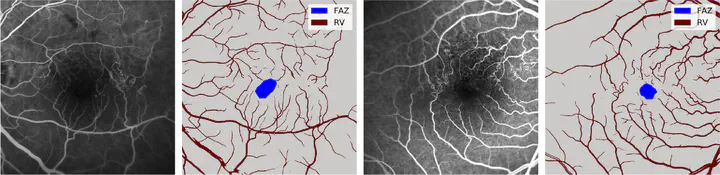
In clinical routine, ophthalmologists frequently analyze the shape and size of the foveal avascular zone (FAZ) to detect and monitor retinal diseases. In order to extract those parameters, the contours of the FAZ need to be segmented, which is normally achieved by analyzing the retinal vasculature (RV) around the macula in fluorescein angiograms (FA). Computer-aided segmentation methods based on deep learning (DL) can automate this task. However, current approaches for segmenting the FAZ are often tailored to a specific dataset or require manual initialization. Furthermore, they do not take the variability and challenges of clinical FA into account, which are often of low quality and difficult to analyze. In this paper we propose a DL-based framework to automatically segment the FAZ in challenging FA scans from clinical routine. Our approach mimics the workflow of retinal experts by using additional RV labels as a guidance during training. Hence, our model is able to produce RV segmentations simultaneously. We minimize the annotation work by using a multi-modal approach that leverages already available public datasets of color fundus pictures (CFPs) and their respective manual RV labels. Our experimental evaluation on two datasets with FA from 1) clinical routine and 2) large multicenter clinical trials shows that the addition of weak RV labels as a guidance during training improves the FAZ segmentation significantly with respect to using only manual FAZ annotations.